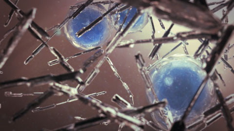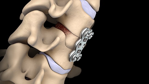

This 3D medical animation shows the normal anatomy of the cervical spine and age-related wear and tear that narrows the vertebral canal. An anterior cervical discectomy and fusion procedure to relieve pressure on the spinal cord is also shown.
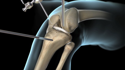

The anterior cruciate ligament, or ACL, is one of the four main ligaments connecting the thighbone to the shinbone. This video shows a surgical procedure to repair a damaged ACL.
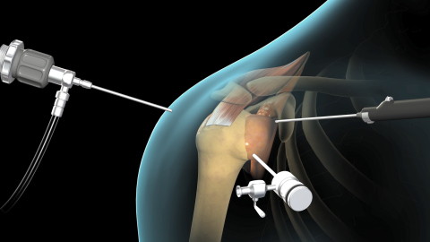

The rotator cuff is a group of arm muscles and tendons that surround the shoulder joint. This video shows a surgical procedure to repair a damaged rotator cuff.
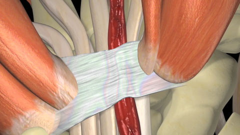

Carpal tunnel syndrome is a disorder caused by compression of a nerve in the wrist. This video shows two different surgical procedures to relieve the nerve compression.
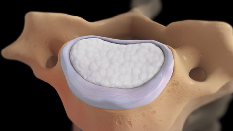

This video shows the anatomy of the spine in the neck and common injuries in this area.
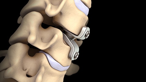

This 3D medical animation shows the normal anatomy of the cervical spine, along with age-related wear and tear that narrow the spinal canal. A cervical disc replacement procedure to relieve compression of the spinal cord and nerves is also show.
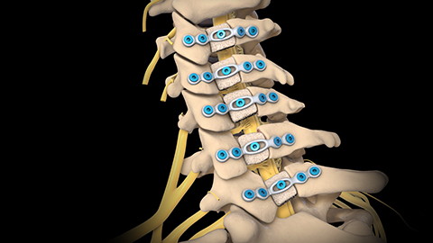

This 3D medical animation shows the normal anatomy of the cervical spine and age-related wear and tear that narrows the vertebral canal. A cervical laminoplasty procedure to relieve pressure on the spinal cord is also shown.


General anesthesia is a treatment used to block pain and put the body to sleep during surgical procedures. This video shows the different ways doctors give patients general anesthesia before a surgical procedure.
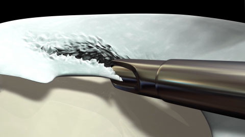

Knee arthroscopy is a surgical procedure to view or repair damage inside the knee through tiny incisions using a tube-like instrument, called an arthroscope, and surgical tools. This video shows the anatomy of the knee and how a surgeon performs a knee arthroscopy.
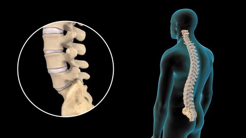

This video shows the anatomy of the spine in the lower back, common injuries in this area, and their treatment.
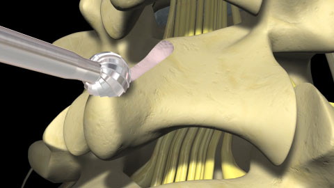

Lumbar laminectomy is a surgical procedure in which a surgeon removes the back of the some of the bones in the spine called vertebrae. The surgeon may also remove parts of the disc, which is the cushioning tissue between each vertebrae.
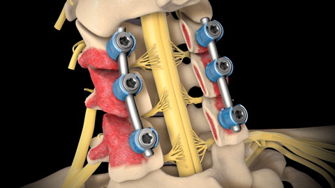

This 3D medical animation shows the normal anatomy of the cervical spine and age-related wear and tear that narrows the vertebral canal. A posterior cervical laminectomy and fusion procedure to relieve pressure on the spinal cord and stabilize the spine is also shown.
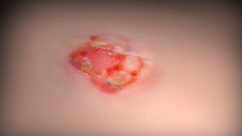

Pressure sores, also called bedsores or decubitus ulcers, are skin injuries caused by sitting or lying in one position too long. This video shows the stages and treatment of pressure sores.
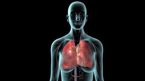

Deep vein thrombosis (DVT) is a condition in which a thrombus, or blood clot, forms on a valve inside a vein, blocking the flow of blood through the vein. This video shows how a thrombus forms and what happens if the thrombus breaks free from the valve.
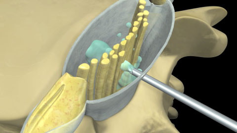

Spinal and epidural anesthesia are injections of liquid drugs into the area surrounding the spinal cord to cause numbness in an area of the body.
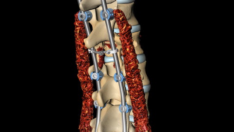

Spinal fusion is a surgical procedure in which a surgeon fuses two or more bones of the spine together. This video shows how and why surgeons perform this procedure.
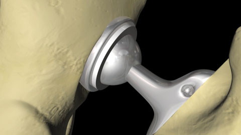

Total hip replacement is a surgical procedure in which the surgeon replaces a damaged, or diseased hip joint with a man-made hip joint. This video shows an open procedure on the back of the hip.


Total hip replacement is a surgical procedure in which the surgeon replaces a damaged or diseased hip joint with a man-made hip joint. This video shows a minimally invasive approach on the front of the hip.
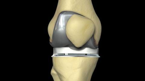

Total knee joint replacement is a surgical procedure to replace a damaged knee joint with a man-made knee joint. This video shows how and why surgeons perform this procedure.
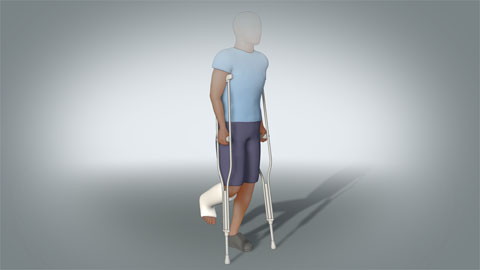

This 3D medical animation details the proper techniques for sitting, standing and walking with crutches following surgery or injury. The views of this animation will learn how to sit, stand and walk with crutches after surgery.
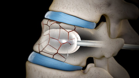

This 3D medical animation shows the normal anatomy of a vertebra in the spine. A compression type fracture of the vertebral body is depicted. Two procedures to repair and stabilize a compression fracture are also shown.
